Moyamoya disease: What to know about this rare brain condition
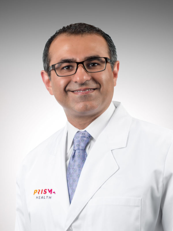
Moyamoya disease is a rare condition caused by blocked arteries in the brain that lead to stroke-like symptoms. Neurosurgeon Roham Moftakhar, MD, explained what causes moyamoya disease, how it’s diagnosed and what treatments are available.
What causes moyamoya disease?
Moyamoya disease is a genetic disease where the carotid arteries in the brain become narrow over time. To try to make up for the lack of blood flow in the brain, small blood vessels called collaterals, or moyamoya vessels, form over time. The term moyamoya is Japanese for “puff of smoke.”
“There are all these very tiny vessels that are coming off of the carotid artery, and they resemble a puff of smoke,” explained Dr. Moftakhar.
What are the symptoms of moyamoya disease?
Symptoms can vary, but moyamoya mainly presents as stroke-like symptoms, which can include:
- A drooping face, mainly on one side
- Weakness in the arms or legs, mainly on one side
- Numbness on the arms or legs, mainly on one side
- Speech problems, such as inability to speak or understand what is said to them
“It doesn’t have to be all of these signs,” said Dr. Moftakhar. “It could also be one or two of them. It varies depending on where the problem is occurring in the brain.”
When these symptoms appear, they usually go away in less than 24 hours. But symptoms can persist longer, and in that case the patient has likely had a full-on stroke.
Are there risk factors for moyamoya?
Moyamoya disease primarily affects children, but it can also occur in adults. It’s more common among people with East Asian heritage, especially Korean, Japanese and Chinese. Having a family history of the disease increases your risk, as well as having other medical conditions such as sickle cell anemia and Down syndrome.
How is moyamoya disease typically found?
Moyamoya is sometimes found by accident in a person with no symptoms. For example, if someone is brought to the emergency room after a fall and a scan of the head could reveals the carotid arteries have moyamoya. However, most patients are diagnosed because of stroke-like symptoms.
If moyamoya disease is suspected, the diagnosis is made using various imaging studies, the first being a CAT scan angiogram which looks at the blood vessels in the brain. If the symptoms are caused by moyamoya, the scan will reveal that there are tiny moyamoya blood vessels, and the internal carotid artery is not healthy.
The next step would be a cerebral angiogram, which is a somewhat invasive but extremely important test. “This test is done by a neurosurgeon, a neuro-interventional radiologist or a neurologist,” explained Dr. Moftakhar. “During this test, we go through the blood vessel with a catheter all the way to the carotid arteries in the neck. From there, we inject dye while taking X-ray pictures. The advantage of a cerebral angiogram is that it’s a dynamic study, meaning that we could look at the different phases of the blood vessels in the brain. This gives us lots of information.”
The cerebral angiogram has a small degree of risk to it, including blood vessel injury, contrast allergy and kidney issues.
The next test used for diagnosis is an MRI of the brain, looking for any type of permanent stroke. “With an MRI, we can see whether the patient has had a permanent stroke, or if this just a transient ischemic attack – a TIA, also known as a mini-stroke,” Dr. Moftakhar said.
If moyamoya is found on both sides of the brain, it’s referred to as moyamoya disease. Moyamoya on one side of the brain only is called moyamoya syndrome.
How is moyamoya treated?
Moyamoya can be treated with medicine, which usually involves taking aspirin. “The medication is not a cure, but it is a way to hopefully improve the process,” Dr. Moftakhar said.
Another option is bypass surgery, of which there are two types – direct and indirect.
Direct bypass surgery for the brain involves using a blood vessel from underneath the scalp called a superficial temporal artery. “If you put two fingers in front of your ear, you can feel the superficial temporal artery pulsating,” Dr. Moftakhar said.
That artery is sewn into one of the blood vessels in the brain, which immediately (and over time) gives the extra blood flow that is needed to the brain.
Indirect bypass surgery for the brain involves taking the superficial temporal artery and laying it on the brain. Since the brain is starved for blood flow, and there are factors that are being produced in the brain to make new blood vessels, the harvested superficial temporal artery that is laid on the brain branches out, creating new blood vessels in about three to six months.
“An advantage of the direct bypass is there can be an immediate supply of blood flow since it’s sewn into the blood vessel in the brain,” Dr. Moftakhar said. “The indirect bypass doesn’t give you immediate blood flow, but it does make new blood vessels over time. Both surgeries are very acceptable, and they’re good solutions for someone with moyamoya who is having symptoms of a stroke or transient ischemic attack.”
Surgeries can take two to five hours. They are done under general anesthesia where the patient is completely asleep, and after the surgery they are carefully watched in a neurological intensive care unit.
The hospital stay is, on average, about three to four days. Total recovery takes between two to six weeks, depending on the surgery and how the patient is doing neurologically.
In patients with moyamoya disease affecting both sides of the brain, the surgery can be done on both sides, but most often there is a gap between the first surgery and the second surgery.
What happens after the bypass surgery?
“After the surgery, the patient must be sure to stay hydrated, maintain a normal blood pressure and take their aspirin,” Dr. Moftakhar said. “This is a critical period because there isn’t an immediate supply of blood flow. So, we have to make sure that the patient keeps hydrated.”
Six months after the surgery, another cerebral angiogram is performed to confirm the surgery has worked and new blood vessels that have formed. After another six months, patients might undergo another CAT scan-based angiogram or another formal cerebral angiogram depending on what their first test has shown.
Patients will continue to receive follow-up checks over their lifetime.
Find a doctor
Whether you’re looking for a primary care physician or need to see a specialist, we’re here to help with experienced, compassionate care near you.
Find a Doctor

