
What kinds of imaging methods does a radiologist use to check for heart problems?

MRI, CTA, calcium scoring … when you’re already worried about what’s happening with your heart, these technical terms and acronyms can make you feel even more confused and stressed. Cardiac imaging tests help doctors discover the underlying reasons for your symptoms, but what exactly are they looking for?
Diagnostic radiologist Pal Suranyi, MD, spoke with us about the kinds of imaging used to check for heart problems, the differences between a CT and an MRI and more.
What are the different types of imaging that might be used to check your heart?
“There are several types of imaging that are used by radiologists when we’re looking into different symptoms or potential conditions,” said Dr. Suranyi. “Some conditions may be more visible using an MRI machine than a cardiac CTA, for instance.”
Types of cardiac imaging commonly used include:
- Coronary calcium scoring: Non-contrast chest CT, sometimes called a CAT scan. Helpful for looking into the risk of coronary disease when a patient doesn’t have symptoms but does have risk factors like family history.
- Coronary CTA, or computed tomographic angiography: Looks into the vessels (coronary arteries) that feed that heart. Used both when there are symptoms like shortness of breath or chest pain but also used in preventive or even primary care. Helpful when working with patients who have mild or unusual symptoms and who have low to moderate risk of disease, but it’s also used for those who are high risk but have no symptoms. Early detection using a coronary CTA helps to slow disease progression, since it could lead to medication changes or help identify disease early, resulting in better treatment outcomes.
- Cardiac (heart) CTA: Used when detecting heart problems like valve disease or looking for masses, clots or other problems within the chambers of the heart. Congenital heart diseases, those you are born with like atrial or ventricular septal defects, can also be detected with this scan.
- Cardiac MRI (magnetic resonance imaging): Often considered the gold standard for testing heart function and valve function when it comes to heart disease, thanks to how well it allows your treatment team to investigate your heart in detail. Patients with heart failure or irregular, fast or skipped beats can benefit from this. Also, those who have had heart attacks before may need a cardiac MRI to help guide treatment.
“I also want to include the chest MRA, or magnetic resonance angiography,” said Dr. Suranyi. “This study is also performed in an MRI scanner. While this test isn’t directly checking on the heart, it does help to check on the blood vessels closely connected to it. For example, a chest MRA can help check your aorta for enlargement without the need for X-ray radiation.”
Chest MRAs help when it comes to screening for aneurysms or monitoring known aortic aneurysms for concerning features that might require intervention. Patients with a history of smoking or high blood pressure are at increased risk for aortic aneurysm or aortic dissection, but family history or genetic or congenital conditions can also increase risk, including conditions like Marfan Syndrome, Ehlers-Danlos syndrome or bicuspid aortic valve.
What kinds of symptoms might lead a medical provider to recommend cardiac imaging?
“The first main symptom you might think of would be typical or unusual chest pain, but there are many other symptoms that could suggest a serious heart problem that can be found through cardiac imaging,” said Dr. Suranyi. “People who are recommended for imaging may be dealing with jaw pain, pain or discomfort in the abdomen, just around the bottom of the ribs, back pain or pain that seems to radiate to the left arm.”
Other common symptoms that could result in needing cardiac imaging include:
- Shortness of breath
- Difficult or heavy breathing during regular activities
- Feeling weak
- Swelling in the legs or feet
- Unusual heart ‘sounds’, such as a new heart murmur
- Dizziness and lightheadedness
- Racing heartbeat or the sense that you have ‘skipped beats’ or that the rhythm of your heart is ‘off’
- Fainting or losing consciousness
- Surviving a stroke or TIA that may have resulted from a blood clot or mass in the heart
I’m not having any of those symptoms. Why is my doctor recommending cardiac imaging?
“If you have a lot of risk factors or a strong family history of heart problems, your physician may decide to order cardiac imaging to screen for potential issues,” said Dr. Suranyi. “For example, if you undergo a mammogram or a screening for lung cancer, it’s possible that there were signs in those unrelated tests that suggest you’re at a higher risk for coronary disease.”
Patients who have had a CT scan after traumatic injury, after fighting serious pneumonia or who have a suspected pulmonary embolism, may have heart findings on those scans that prompt your doctors to send you for a CTA to rule out coronary disease as well.
“’Silent heart attacks and other heart conditions, that just don’t present with the symptoms we’re all told to watch out for, are very real,” added Dr. Suranyi. “Even if you’re not sure why the test was ordered, it can often discover problems that otherwise might be missed.”
What are medical providers looking for when they use cardiac imaging?
“Generally, we’re looking to learn about the heart chamber sizes, how well it’s functioning, how thick the muscle of your heart is and its overall performance, appearance and function of the valves and the overall conditions of the vessels around the heart,” said Dr. Suranyi.
Your doctor may also want to find out if there are abnormal masses in the heart or check for potentially dangerous blood clots. Cardiac imaging has an important role in planning transcatheter interventions, surgeries or potentially implanting devices like artificial valves. During follow up, cardiac imaging helps assess results and catch and treat complications.
How is imaging used when preparing for, during immediate treatment or during follow-ups afterward?
“We use imaging to get precise measurements of the structures in the heart and vessels,” said Dr. Suranyi. “It helps your doctors select the treatment plan that works for each individual patient. We also can get a closer look at how well blood is flowing to and from the heart, which helps with timing of procedures.”
At later follow-up appointments, imaging helps to measure improvement and check device position. It also helps your care team to find potential complications.
What is the difference between a cardiac CT and an MRI?
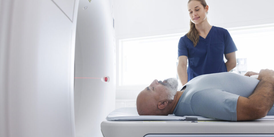
Cardiac computed tomography, or cardiac CT, can be visualized as basically a big spinning donut with an X-ray source on one side and X-ray detectors on the other. The device takes many X-rays around the body quite quickly and helps us get an accurate visual. Since the heart is always beating, the image acquisition has to be timed to your heartbeat (ECG). You have to hold still and hold your breath for 10-15 seconds, while we are taking pictures, as the table moves you through the donut.
“A cardiac MRI is still best visualized as a donut, although this one is more of a tube shape. It’s a large magnet that doesn’t utilize any X-ray radiation at all. Instead, it generates gentle microwaves,” Dr. Suranyi added. “We use these microwaves to spin the water and other molecules around in your body, then we record the signal that comes from them using an ‘antenna,’ a coil placed on your chest. You will also have ECG electrodes placed on your chest, so that the imaging can be timed to the beating of your heart.”
Patients undergoing an MRI usually must be still for about 30-45 minutes, depending on what your care team is looking for. You’ll be asked to briefly hold your breath for 10-15 seconds at a time multiple times during the exam.
For both CTA and MRI, you might be given a ‘contrast dye’ through an IV to help visualize the inside of vessels and your heart more clearly.
Why choose cardiac CT?
“CT scans are very quick and give us excellent resolution on images of the heart and its vessels,” said Dr. Suranyi. “It’s fairly noninvasive and can be done quickly in outpatient or inpatient settings, so it’s very adaptable to each patient’s circumstances. There’s some prep involved, since there’s an IV line and ECG electrodes and some time is needed to check on vitals and slow down your heartrate, but the actual scan only takes about 15 seconds.”
CT scans are also far more widely available than an MRI, and results come back more quickly.
Why choose a cardiac MRI?
“There is simply no other imaging test with the same ability to visualize diseases of the heart muscle at this level,” said Dr. Suranyi. “MRI testing detects subtle, early disease and changes in the heart, sometimes even before they affect function or rhythm, and provides the most accurate measurements of heart function, muscle thickness and chamber sizes.”
An MRI takes longer and is more demanding on the patient than a cardiac CT, as well as not being available at every facility.
Why are noninvasive imaging methods recommended for heart tests?
“These noninvasive testing methods are quicker, easier to schedule and carry less risk than the more invasive cardiac evaluations,” said Dr. Suranyi.
Noninvasive tests can also offer alternative explanations for symptoms that mimic heart disease, such as chest pain, that end up being caused by other conditions entirely.
What kind of preparation is there for these cardiac imaging methods?
“Generally, we advise no caffeine for 12 hours prior to testing, avoid taking any erectile dysfunction medicines for 48 hours before your CTA scan, and, if possible, avoid using inhalers for asthma,” said Dr. Suranyi. “If you need to use your inhaler in that time period, just make sure you let your nurses, technologists or doctors know.”
If you have any implanted devices, bring the documentation on your device with you to the MRI scanner to make sure they can be reviewed for safety.
What’s your heart age?
Take a free, fast health assessment and see if you’re at risk for heart disease.
Start Now